Calcium »
PDB 7brw-7c4h »
7bsf »
Calcium in PDB 7bsf: Crystal Structure of A Nir-Emitting Dna-Stabilized AG16 Nanocluster (T5C Mutant)
Protein crystallography data
The structure of Crystal Structure of A Nir-Emitting Dna-Stabilized AG16 Nanocluster (T5C Mutant), PDB code: 7bsf
was solved by
J.Kondo,
C.Cerretani,
T.Vosch,
with X-Ray Crystallography technique. A brief refinement statistics is given in the table below:
| Resolution Low / High (Å) | 28.43 / 1.10 |
| Space group | P 21 21 21 |
| Cell size a, b, c (Å), α, β, γ (°) | 23.958, 33.807, 52.523, 90.00, 90.00, 90.00 |
| R / Rfree (%) | 7.5 / 8.3 |
Other elements in 7bsf:
The structure of Crystal Structure of A Nir-Emitting Dna-Stabilized AG16 Nanocluster (T5C Mutant) also contains other interesting chemical elements:
| Silver | (Ag) | 18 atoms |
Calcium Binding Sites:
The binding sites of Calcium atom in the Crystal Structure of A Nir-Emitting Dna-Stabilized AG16 Nanocluster (T5C Mutant)
(pdb code 7bsf). This binding sites where shown within
5.0 Angstroms radius around Calcium atom.
In total only one binding site of Calcium was determined in the Crystal Structure of A Nir-Emitting Dna-Stabilized AG16 Nanocluster (T5C Mutant), PDB code: 7bsf:
In total only one binding site of Calcium was determined in the Crystal Structure of A Nir-Emitting Dna-Stabilized AG16 Nanocluster (T5C Mutant), PDB code: 7bsf:
Calcium binding site 1 out of 1 in 7bsf
Go back to
Calcium binding site 1 out
of 1 in the Crystal Structure of A Nir-Emitting Dna-Stabilized AG16 Nanocluster (T5C Mutant)
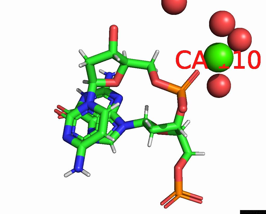
Mono view
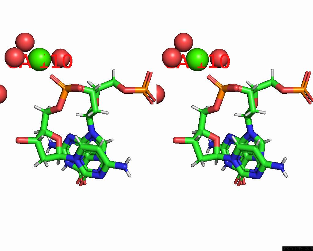
Stereo pair view
Mono view
Stereo pair view
A full contact list of Calcium with other atoms in the Ca binding
site number 1 of Crystal Structure of A Nir-Emitting Dna-Stabilized AG16 Nanocluster (T5C Mutant) within 5.0Å range:
|
Reference:
C.Cerretani,
J.Kondo,
T.Vosch.
Mutation of Position 5 As A Crystal Engineering Tool For A Nir-Emitting Dna-Stabilized AG16 Nanocluster. Crystengcomm V. 22 8136 2020.
ISSN: ISSN 1466-8033
DOI: 10.1039/D0CE01225D
Page generated: Thu Jul 18 23:22:01 2024
ISSN: ISSN 1466-8033
DOI: 10.1039/D0CE01225D
Last articles
Zn in 9MJ5Zn in 9HNW
Zn in 9G0L
Zn in 9FNE
Zn in 9DZN
Zn in 9E0I
Zn in 9D32
Zn in 9DAK
Zn in 8ZXC
Zn in 8ZUF