Calcium »
PDB 7brx-7c52 »
7bys »
Calcium in PDB 7bys: Crystal Structure of Exo-Beta-1,3-Galactanase From Phanerochaete Chrysosporium PC1,3GAL43A Apo Form
Enzymatic activity of Crystal Structure of Exo-Beta-1,3-Galactanase From Phanerochaete Chrysosporium PC1,3GAL43A Apo Form
All present enzymatic activity of Crystal Structure of Exo-Beta-1,3-Galactanase From Phanerochaete Chrysosporium PC1,3GAL43A Apo Form:
3.2.1.145;
3.2.1.145;
Protein crystallography data
The structure of Crystal Structure of Exo-Beta-1,3-Galactanase From Phanerochaete Chrysosporium PC1,3GAL43A Apo Form, PDB code: 7bys
was solved by
K.Matsuyama,
T.Ishida,
N.Kishine,
Z.Fujimoto,
K.Igarashi,
S.Kaneko,
with X-Ray Crystallography technique. A brief refinement statistics is given in the table below:
| Resolution Low / High (Å) | 8.00 / 1.40 |
| Space group | P 1 |
| Cell size a, b, c (Å), α, β, γ (°) | 40.470, 66.262, 74.021, 72.03, 84.68, 82.15 |
| R / Rfree (%) | 15.5 / 18.6 |
Calcium Binding Sites:
The binding sites of Calcium atom in the Crystal Structure of Exo-Beta-1,3-Galactanase From Phanerochaete Chrysosporium PC1,3GAL43A Apo Form
(pdb code 7bys). This binding sites where shown within
5.0 Angstroms radius around Calcium atom.
In total 2 binding sites of Calcium where determined in the Crystal Structure of Exo-Beta-1,3-Galactanase From Phanerochaete Chrysosporium PC1,3GAL43A Apo Form, PDB code: 7bys:
Jump to Calcium binding site number: 1; 2;
In total 2 binding sites of Calcium where determined in the Crystal Structure of Exo-Beta-1,3-Galactanase From Phanerochaete Chrysosporium PC1,3GAL43A Apo Form, PDB code: 7bys:
Jump to Calcium binding site number: 1; 2;
Calcium binding site 1 out of 2 in 7bys
Go back to
Calcium binding site 1 out
of 2 in the Crystal Structure of Exo-Beta-1,3-Galactanase From Phanerochaete Chrysosporium PC1,3GAL43A Apo Form
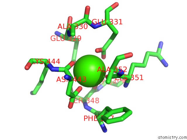
Mono view
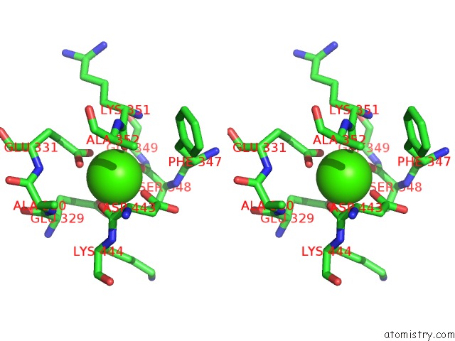
Stereo pair view
Mono view
Stereo pair view
A full contact list of Calcium with other atoms in the Ca binding
site number 1 of Crystal Structure of Exo-Beta-1,3-Galactanase From Phanerochaete Chrysosporium PC1,3GAL43A Apo Form within 5.0Å range:
|
Calcium binding site 2 out of 2 in 7bys
Go back to
Calcium binding site 2 out
of 2 in the Crystal Structure of Exo-Beta-1,3-Galactanase From Phanerochaete Chrysosporium PC1,3GAL43A Apo Form
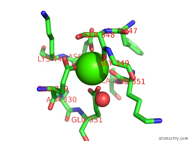
Mono view
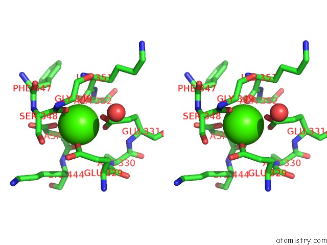
Stereo pair view
Mono view
Stereo pair view
A full contact list of Calcium with other atoms in the Ca binding
site number 2 of Crystal Structure of Exo-Beta-1,3-Galactanase From Phanerochaete Chrysosporium PC1,3GAL43A Apo Form within 5.0Å range:
|
Reference:
K.Matsuyama,
N.Kishine,
Z.Fujimoto,
N.Sunagawa,
T.Kotake,
Y.Tsumuraya,
M.Samejima,
K.Igarashi,
S.Kaneko.
Unique Active Site and Subsite Features in the Arabinogalactan-Degrading GH43 Exo-Beta-1,3-Galactanase From Phanerochaete Chrysosporium J.Biol.Chem. 2020.
ISSN: ESSN 1083-351X
PubMed: 33093171
DOI: 10.1074/JBC.RA120.016149
Page generated: Wed Jul 9 21:04:22 2025
ISSN: ESSN 1083-351X
PubMed: 33093171
DOI: 10.1074/JBC.RA120.016149
Last articles
Cl in 5J2DCl in 5J2C
Cl in 5J23
Cl in 5J2B
Cl in 5J2A
Cl in 5IXL
Cl in 5J0Z
Cl in 5J1T
Cl in 5J1S
Cl in 5IXY