Calcium »
PDB 7pod-7qfy »
7psp »
Calcium in PDB 7psp: Crystal Structure of S100A4 Labeled with NU000846B.
Protein crystallography data
The structure of Crystal Structure of S100A4 Labeled with NU000846B., PDB code: 7psp
was solved by
C.Giroud,
T.Szommer,
C.Coxon,
O.Monteiro,
T.Christott,
J.Bennett,
K.Aitmakhanova,
B.Raux,
J.Newman,
J.Elkins,
G.Arruda Bezerra,
T.Krojer,
L.Koekemoer,
F.Von Delft,
C.Bountr,
P.Brennan,
O.Fedorov,
with X-Ray Crystallography technique. A brief refinement statistics is given in the table below:
| Resolution Low / High (Å) | 54.77 / 2.61 |
| Space group | P 64 2 2 |
| Cell size a, b, c (Å), α, β, γ (°) | 109.54, 109.54, 89.87, 90, 90, 120 |
| R / Rfree (%) | 24.5 / 28.6 |
Other elements in 7psp:
The structure of Crystal Structure of S100A4 Labeled with NU000846B. also contains other interesting chemical elements:
| Chlorine | (Cl) | 2 atoms |
Calcium Binding Sites:
The binding sites of Calcium atom in the Crystal Structure of S100A4 Labeled with NU000846B.
(pdb code 7psp). This binding sites where shown within
5.0 Angstroms radius around Calcium atom.
In total 4 binding sites of Calcium where determined in the Crystal Structure of S100A4 Labeled with NU000846B., PDB code: 7psp:
Jump to Calcium binding site number: 1; 2; 3; 4;
In total 4 binding sites of Calcium where determined in the Crystal Structure of S100A4 Labeled with NU000846B., PDB code: 7psp:
Jump to Calcium binding site number: 1; 2; 3; 4;
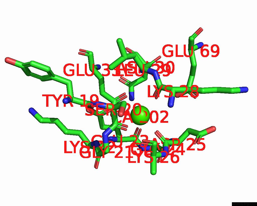
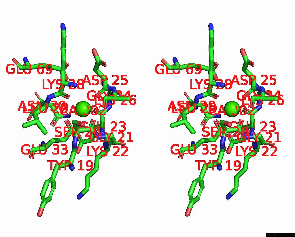
Calcium binding site 1 out of 4 in 7psp
Go back to
Calcium binding site 1 out
of 4 in the Crystal Structure of S100A4 Labeled with NU000846B.
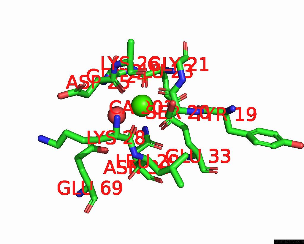
Mono view
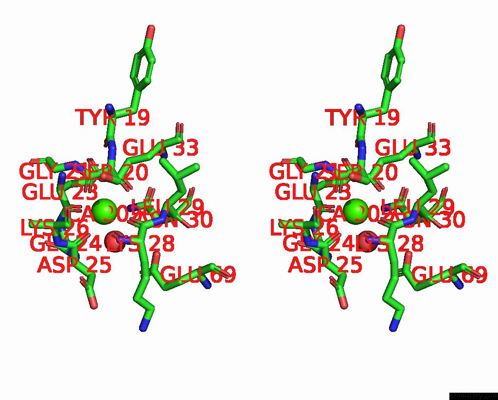
Stereo pair view
Mono view
Stereo pair view
A full contact list of Calcium with other atoms in the Ca binding
site number 1 of Crystal Structure of S100A4 Labeled with NU000846B. within 5.0Å range:
|
Calcium binding site 2 out of 4 in 7psp
Go back to
Calcium binding site 2 out
of 4 in the Crystal Structure of S100A4 Labeled with NU000846B.
Mono view

Stereo pair view
Mono view
Stereo pair view
A full contact list of Calcium with other atoms in the Ca binding
site number 2 of Crystal Structure of S100A4 Labeled with NU000846B. within 5.0Å range:
|
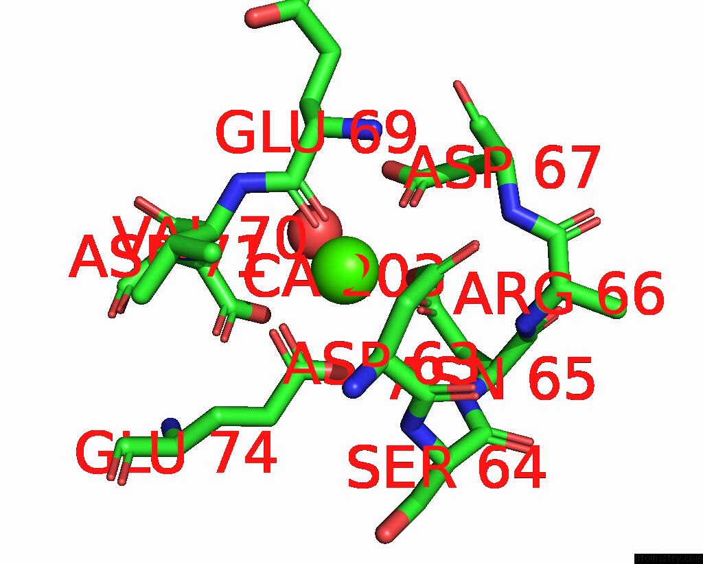
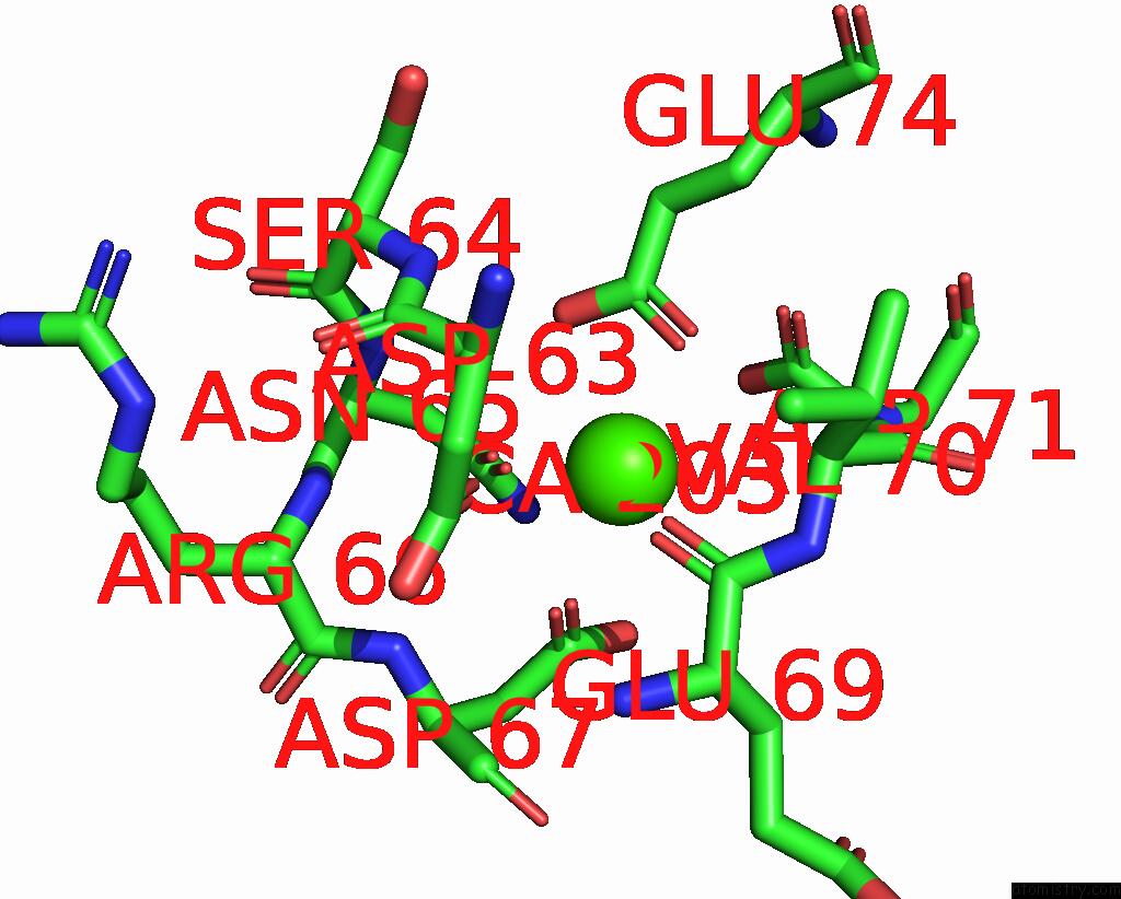
Calcium binding site 3 out of 4 in 7psp
Go back to
Calcium binding site 3 out
of 4 in the Crystal Structure of S100A4 Labeled with NU000846B.
Mono view
Stereo pair view
Mono view
Stereo pair view
A full contact list of Calcium with other atoms in the Ca binding
site number 3 of Crystal Structure of S100A4 Labeled with NU000846B. within 5.0Å range:
|
Calcium binding site 4 out of 4 in 7psp
Go back to
Calcium binding site 4 out
of 4 in the Crystal Structure of S100A4 Labeled with NU000846B.
Mono view

Stereo pair view
Mono view
Stereo pair view
A full contact list of Calcium with other atoms in the Ca binding
site number 4 of Crystal Structure of S100A4 Labeled with NU000846B. within 5.0Å range:
|
Reference:
C.Giroud,
C.Giroud,
T.Szommer,
C.Coxon,
O.Monteiro,
T.Christott,
J.Bennett,
K.Aitmakhanova,
B.Raux,
J.Newman,
J.Elkins,
G.Arruda Bezerra,
T.Krojer,
L.Koekemoer,
F.Von Delft,
C.Bountr,
P.Brennan,
O.Fedorov.
N/A N/A.
Page generated: Thu Jul 10 00:23:17 2025
Last articles
Fe in 2YXOFe in 2YRS
Fe in 2YXC
Fe in 2YNM
Fe in 2YVJ
Fe in 2YP1
Fe in 2YU2
Fe in 2YU1
Fe in 2YQB
Fe in 2YOO