Calcium »
PDB 7pod-7qfy »
7pvn »
Calcium in PDB 7pvn: Crystal Structure of Human UBA6 in Complex with Atp
Enzymatic activity of Crystal Structure of Human UBA6 in Complex with Atp
All present enzymatic activity of Crystal Structure of Human UBA6 in Complex with Atp:
6.2.1.45;
6.2.1.45;
Protein crystallography data
The structure of Crystal Structure of Human UBA6 in Complex with Atp, PDB code: 7pvn
was solved by
N.Truongvan,
S.Li,
H.Schindelin,
with X-Ray Crystallography technique. A brief refinement statistics is given in the table below:
| Resolution Low / High (Å) | 19.98 / 2.71 |
| Space group | C 1 2 1 |
| Cell size a, b, c (Å), α, β, γ (°) | 123.949, 113.748, 183.537, 90, 96.49, 90 |
| R / Rfree (%) | 23.3 / 26.2 |
Other elements in 7pvn:
The structure of Crystal Structure of Human UBA6 in Complex with Atp also contains other interesting chemical elements:
| Magnesium | (Mg) | 2 atoms |
| Chlorine | (Cl) | 1 atom |
| Arsenic | (As) | 16 atoms |
Calcium Binding Sites:
The binding sites of Calcium atom in the Crystal Structure of Human UBA6 in Complex with Atp
(pdb code 7pvn). This binding sites where shown within
5.0 Angstroms radius around Calcium atom.
In total 3 binding sites of Calcium where determined in the Crystal Structure of Human UBA6 in Complex with Atp, PDB code: 7pvn:
Jump to Calcium binding site number: 1; 2; 3;
In total 3 binding sites of Calcium where determined in the Crystal Structure of Human UBA6 in Complex with Atp, PDB code: 7pvn:
Jump to Calcium binding site number: 1; 2; 3;
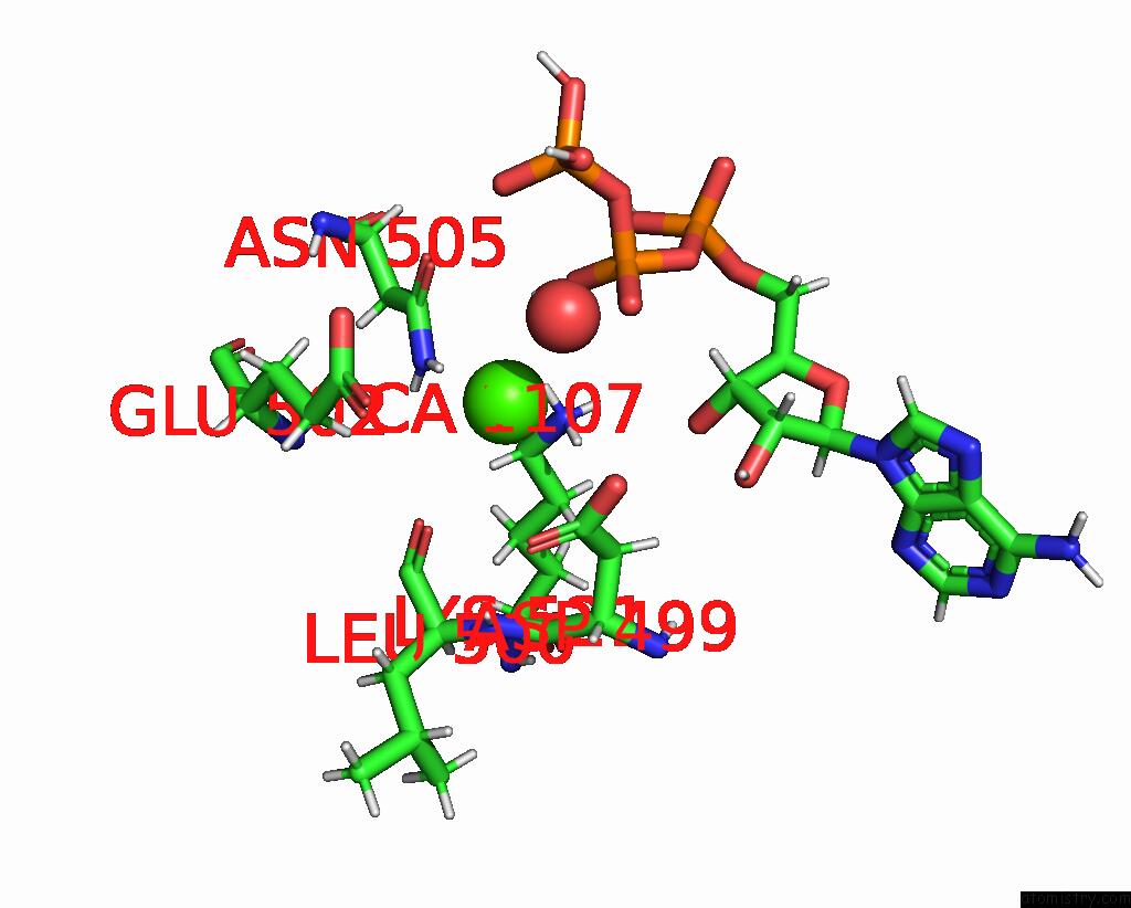
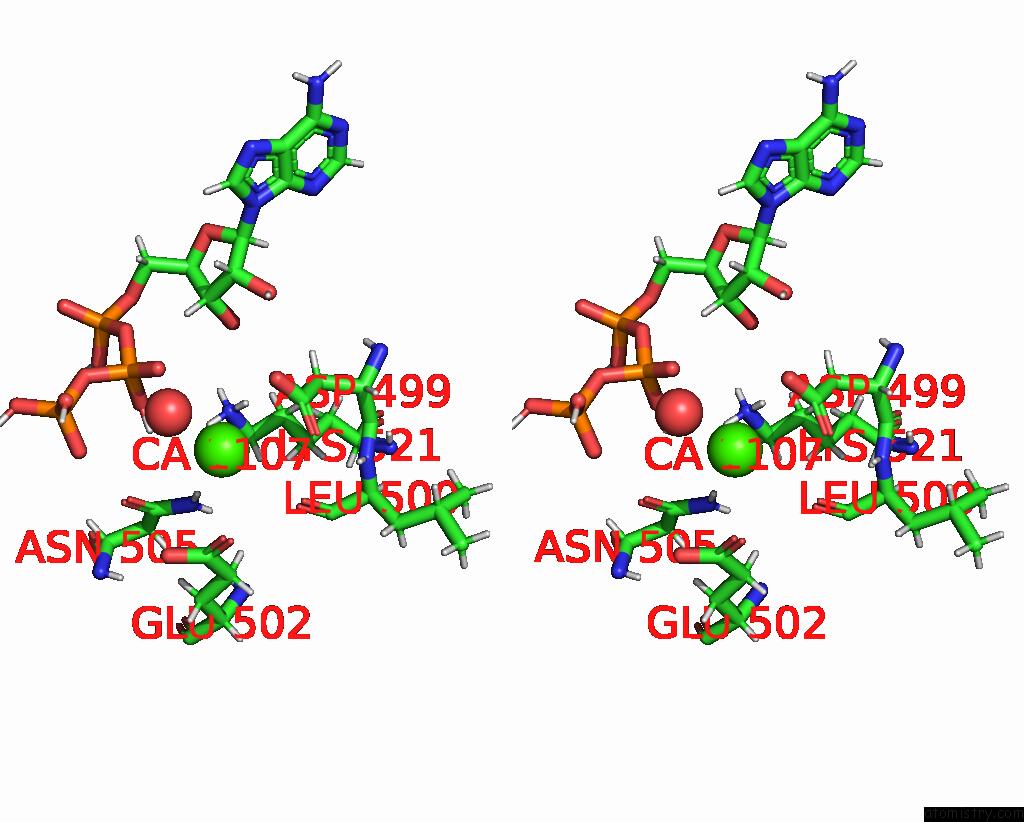
Calcium binding site 1 out of 3 in 7pvn
Go back to
Calcium binding site 1 out
of 3 in the Crystal Structure of Human UBA6 in Complex with Atp
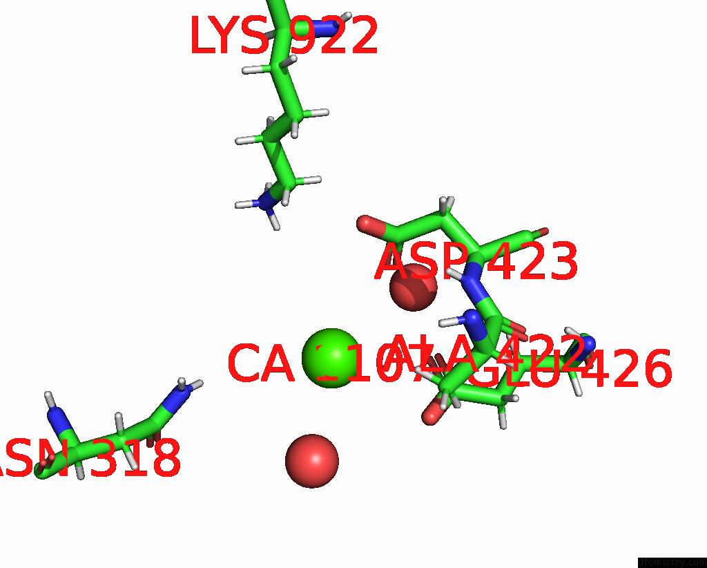
Mono view
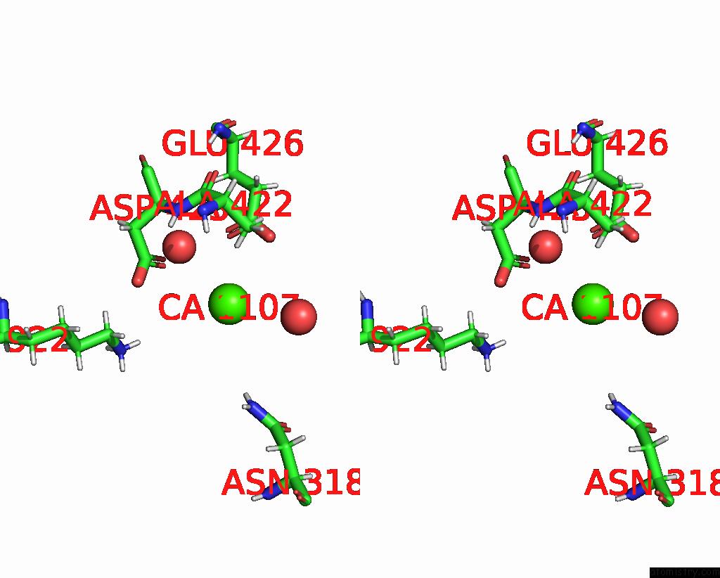
Stereo pair view
Mono view
Stereo pair view
A full contact list of Calcium with other atoms in the Ca binding
site number 1 of Crystal Structure of Human UBA6 in Complex with Atp within 5.0Å range:
|
Calcium binding site 2 out of 3 in 7pvn
Go back to
Calcium binding site 2 out
of 3 in the Crystal Structure of Human UBA6 in Complex with Atp
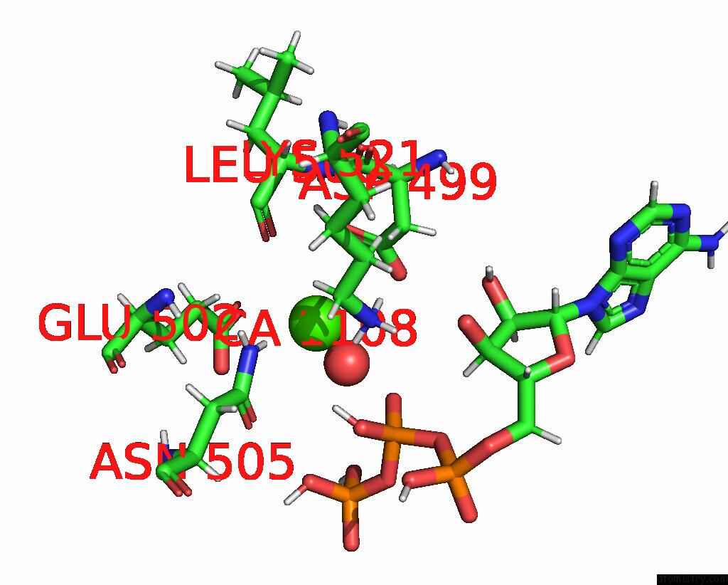
Mono view
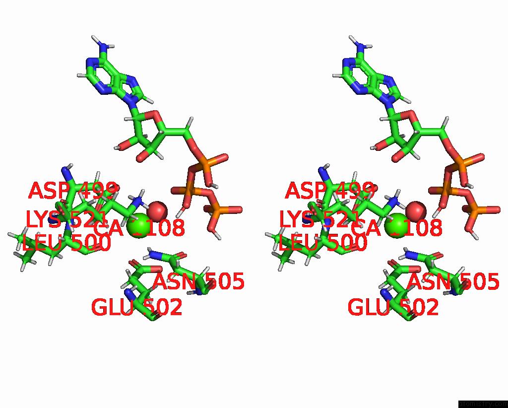
Stereo pair view
Mono view
Stereo pair view
A full contact list of Calcium with other atoms in the Ca binding
site number 2 of Crystal Structure of Human UBA6 in Complex with Atp within 5.0Å range:
|
Calcium binding site 3 out of 3 in 7pvn
Go back to
Calcium binding site 3 out
of 3 in the Crystal Structure of Human UBA6 in Complex with Atp
Mono view
Stereo pair view
Mono view
Stereo pair view
A full contact list of Calcium with other atoms in the Ca binding
site number 3 of Crystal Structure of Human UBA6 in Complex with Atp within 5.0Å range:
|
Reference:
N.Truongvan,
S.Li,
M.Misra,
M.Kuhn,
H.Schindelin.
Structures of UBA6 Explain Its Dual Specificity For Ubiquitin and FAT10. Nat Commun V. 13 4789 2022.
ISSN: ESSN 2041-1723
PubMed: 35970836
DOI: 10.1038/S41467-022-32040-6
Page generated: Thu Jul 10 00:27:37 2025
ISSN: ESSN 2041-1723
PubMed: 35970836
DOI: 10.1038/S41467-022-32040-6
Last articles
Cl in 8DV3Cl in 8DW5
Cl in 8DV4
Cl in 8DU1
Cl in 8DUZ
Cl in 8DTQ
Cl in 8DT6
Cl in 8DSZ
Cl in 8DTE
Cl in 8DTJ