Calcium »
PDB 7pod-7qfy »
7q0x »
Calcium in PDB 7q0x: Bovine Trypsin Co-Crystallized with V(IV)OSO4 and Pic
Enzymatic activity of Bovine Trypsin Co-Crystallized with V(IV)OSO4 and Pic
All present enzymatic activity of Bovine Trypsin Co-Crystallized with V(IV)OSO4 and Pic:
3.4.21.4;
3.4.21.4;
Protein crystallography data
The structure of Bovine Trypsin Co-Crystallized with V(IV)OSO4 and Pic, PDB code: 7q0x
was solved by
M.F.A.Santos,
A.C.P.Fernandes,
I.Correia,
G.Sciortino,
E.Garribba,
T.Santos-Silva,
J.C.Pessoa,
with X-Ray Crystallography technique. A brief refinement statistics is given in the table below:
| Resolution Low / High (Å) | 41.22 / 1.09 |
| Space group | P 31 2 1 |
| Cell size a, b, c (Å), α, β, γ (°) | 54.522, 54.522, 108.199, 90, 90, 120 |
| R / Rfree (%) | 11.1 / 13.6 |
Other elements in 7q0x:
The structure of Bovine Trypsin Co-Crystallized with V(IV)OSO4 and Pic also contains other interesting chemical elements:
| Vanadium | (V) | 1 atom |
Calcium Binding Sites:
The binding sites of Calcium atom in the Bovine Trypsin Co-Crystallized with V(IV)OSO4 and Pic
(pdb code 7q0x). This binding sites where shown within
5.0 Angstroms radius around Calcium atom.
In total only one binding site of Calcium was determined in the Bovine Trypsin Co-Crystallized with V(IV)OSO4 and Pic, PDB code: 7q0x:
In total only one binding site of Calcium was determined in the Bovine Trypsin Co-Crystallized with V(IV)OSO4 and Pic, PDB code: 7q0x:
Calcium binding site 1 out of 1 in 7q0x
Go back to
Calcium binding site 1 out
of 1 in the Bovine Trypsin Co-Crystallized with V(IV)OSO4 and Pic
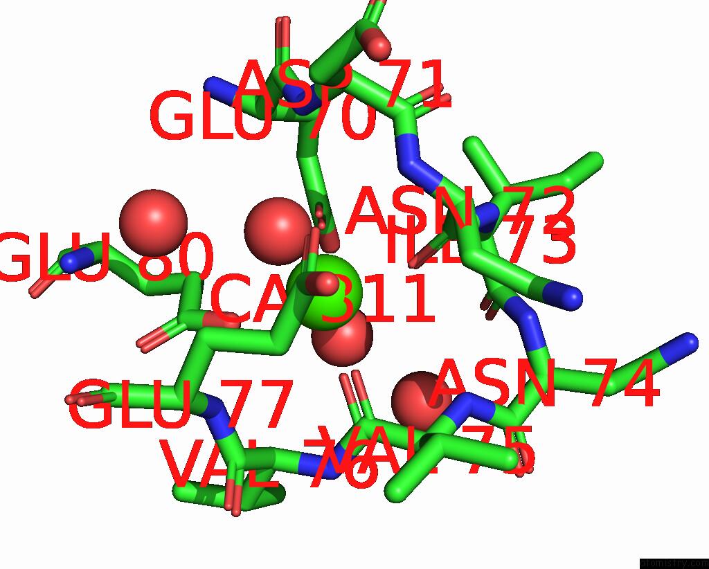
Mono view
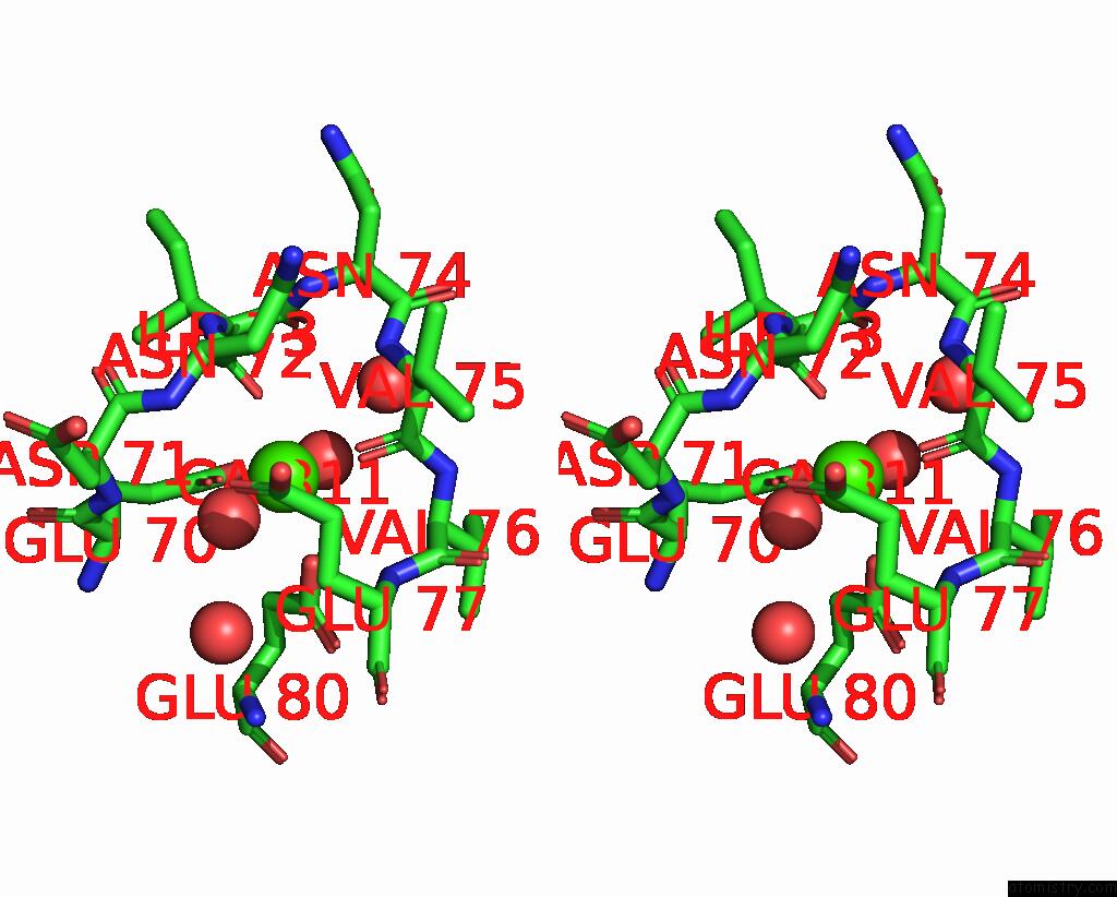
Stereo pair view
Mono view
Stereo pair view
A full contact list of Calcium with other atoms in the Ca binding
site number 1 of Bovine Trypsin Co-Crystallized with V(IV)OSO4 and Pic within 5.0Å range:
|
Reference:
M.F.A.Santos,
G.Sciortino,
I.Correia,
A.C.P.Fernandes,
T.Santos-Silva,
F.Pisanu,
E.Garribba,
J.Costa Pessoa.
Binding of V IV O 2+ , V IV Ol, V IV Ol 2 and V V O 2 L Moieties to Proteins: X-Ray/Theoretical Characterization and Biological Implications. Chemistry V. 28 00105 2022.
ISSN: ISSN 0947-6539
PubMed: 35486702
DOI: 10.1002/CHEM.202200105
Page generated: Thu Jul 10 00:30:32 2025
ISSN: ISSN 0947-6539
PubMed: 35486702
DOI: 10.1002/CHEM.202200105
Last articles
Fe in 2YXOFe in 2YRS
Fe in 2YXC
Fe in 2YNM
Fe in 2YVJ
Fe in 2YP1
Fe in 2YU2
Fe in 2YU1
Fe in 2YQB
Fe in 2YOO