Calcium »
PDB 1dva-1edh »
1dva »
Calcium in PDB 1dva: Crystal Structure of the Complex Between the Peptide Exosite Inhibitor E-76 and Coagulation Factor Viia
Enzymatic activity of Crystal Structure of the Complex Between the Peptide Exosite Inhibitor E-76 and Coagulation Factor Viia
All present enzymatic activity of Crystal Structure of the Complex Between the Peptide Exosite Inhibitor E-76 and Coagulation Factor Viia:
3.4.21.21;
3.4.21.21;
Protein crystallography data
The structure of Crystal Structure of the Complex Between the Peptide Exosite Inhibitor E-76 and Coagulation Factor Viia, PDB code: 1dva
was solved by
C.Eigenbrot,
M.H.Ultsch,
with X-Ray Crystallography technique. A brief refinement statistics is given in the table below:
| Resolution Low / High (Å) | 50.00 / 3.00 |
| Space group | P 1 21 1 |
| Cell size a, b, c (Å), α, β, γ (°) | 70.490, 55.260, 111.730, 90.00, 99.48, 90.00 |
| R / Rfree (%) | 22.5 / 29.5 |
Other elements in 1dva:
The structure of Crystal Structure of the Complex Between the Peptide Exosite Inhibitor E-76 and Coagulation Factor Viia also contains other interesting chemical elements:
| Arsenic | (As) | 2 atoms |
Calcium Binding Sites:
The binding sites of Calcium atom in the Crystal Structure of the Complex Between the Peptide Exosite Inhibitor E-76 and Coagulation Factor Viia
(pdb code 1dva). This binding sites where shown within
5.0 Angstroms radius around Calcium atom.
In total 4 binding sites of Calcium where determined in the Crystal Structure of the Complex Between the Peptide Exosite Inhibitor E-76 and Coagulation Factor Viia, PDB code: 1dva:
Jump to Calcium binding site number: 1; 2; 3; 4;
In total 4 binding sites of Calcium where determined in the Crystal Structure of the Complex Between the Peptide Exosite Inhibitor E-76 and Coagulation Factor Viia, PDB code: 1dva:
Jump to Calcium binding site number: 1; 2; 3; 4;
Calcium binding site 1 out of 4 in 1dva
Go back to
Calcium binding site 1 out
of 4 in the Crystal Structure of the Complex Between the Peptide Exosite Inhibitor E-76 and Coagulation Factor Viia

Mono view
Stereo pair view
Mono view
Stereo pair view
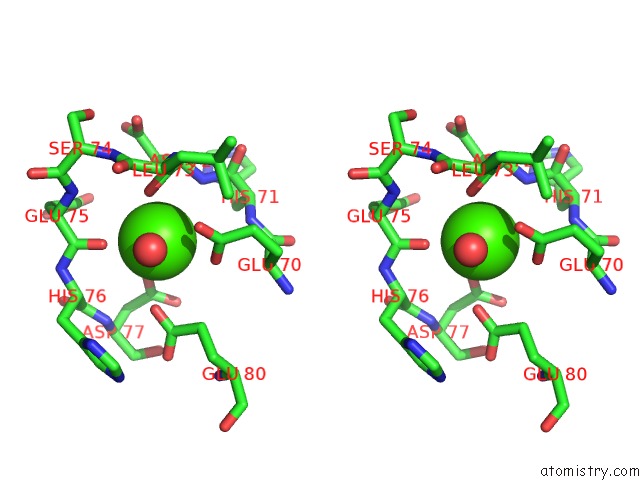
A full contact list of Calcium with other atoms in the Ca binding
site number 1 of Crystal Structure of the Complex Between the Peptide Exosite Inhibitor E-76 and Coagulation Factor Viia within 5.0Å range:
|
Calcium binding site 2 out of 4 in 1dva
Go back to
Calcium binding site 2 out
of 4 in the Crystal Structure of the Complex Between the Peptide Exosite Inhibitor E-76 and Coagulation Factor Viia

Mono view

Stereo pair view
Mono view
Stereo pair view
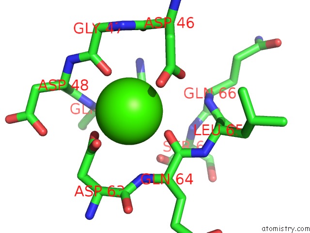
A full contact list of Calcium with other atoms in the Ca binding
site number 2 of Crystal Structure of the Complex Between the Peptide Exosite Inhibitor E-76 and Coagulation Factor Viia within 5.0Å range:
|
Calcium binding site 3 out of 4 in 1dva
Go back to
Calcium binding site 3 out
of 4 in the Crystal Structure of the Complex Between the Peptide Exosite Inhibitor E-76 and Coagulation Factor Viia

Mono view

Stereo pair view
Mono view
Stereo pair view
A full contact list of Calcium with other atoms in the Ca binding
site number 3 of Crystal Structure of the Complex Between the Peptide Exosite Inhibitor E-76 and Coagulation Factor Viia within 5.0Å range:
|
Calcium binding site 4 out of 4 in 1dva
Go back to
Calcium binding site 4 out
of 4 in the Crystal Structure of the Complex Between the Peptide Exosite Inhibitor E-76 and Coagulation Factor Viia
Mono view

Stereo pair view
Mono view
Stereo pair view
A full contact list of Calcium with other atoms in the Ca binding
site number 4 of Crystal Structure of the Complex Between the Peptide Exosite Inhibitor E-76 and Coagulation Factor Viia within 5.0Å range:
|
Reference:
M.S.Dennis,
C.Eigenbrot,
N.J.Skelton,
M.H.Ultsch,
L.Santell,
M.A.Dwyer,
M.P.O'connell,
R.A.Lazarus.
Peptide Exosite Inhibitors of Factor Viia As Anticoagulants. Nature V. 404 465 2000.
ISSN: ISSN 0028-0836
PubMed: 10761907
DOI: 10.1038/35006574
Page generated: Mon Jul 7 14:30:44 2025
ISSN: ISSN 0028-0836
PubMed: 10761907
DOI: 10.1038/35006574
Last articles
I in 6DPHI in 6DPE
I in 6DPF
I in 6DPG
I in 6DPD
I in 6DPC
I in 6DPB
I in 6DPA
I in 6DP9
I in 6DP6