Calcium »
PDB 2rf7-2tep »
2stb »
Calcium in PDB 2stb: Anionic Salmon Trypsin in Complex with Squash Seed Inhibitor (Cucurbita Pepo Trypsin Inhibitor II)
Enzymatic activity of Anionic Salmon Trypsin in Complex with Squash Seed Inhibitor (Cucurbita Pepo Trypsin Inhibitor II)
All present enzymatic activity of Anionic Salmon Trypsin in Complex with Squash Seed Inhibitor (Cucurbita Pepo Trypsin Inhibitor II):
3.4.21.4;
3.4.21.4;
Protein crystallography data
The structure of Anionic Salmon Trypsin in Complex with Squash Seed Inhibitor (Cucurbita Pepo Trypsin Inhibitor II), PDB code: 2stb
was solved by
R.Helland,
G.I.Berglund,
J.Otlewski,
W.Apostoluk,
O.A.Andersen,
N.P.Willassen,
A.O.Smalas,
with X-Ray Crystallography technique. A brief refinement statistics is given in the table below:
| Resolution Low / High (Å) | 6.00 / 1.80 |
| Space group | P 21 21 21 |
| Cell size a, b, c (Å), α, β, γ (°) | 62.300, 63.400, 82.500, 90.00, 90.00, 90.00 |
| R / Rfree (%) | 17.6 / 20.1 |
Calcium Binding Sites:
The binding sites of Calcium atom in the Anionic Salmon Trypsin in Complex with Squash Seed Inhibitor (Cucurbita Pepo Trypsin Inhibitor II)
(pdb code 2stb). This binding sites where shown within
5.0 Angstroms radius around Calcium atom.
In total only one binding site of Calcium was determined in the Anionic Salmon Trypsin in Complex with Squash Seed Inhibitor (Cucurbita Pepo Trypsin Inhibitor II), PDB code: 2stb:
In total only one binding site of Calcium was determined in the Anionic Salmon Trypsin in Complex with Squash Seed Inhibitor (Cucurbita Pepo Trypsin Inhibitor II), PDB code: 2stb:
Calcium binding site 1 out of 1 in 2stb
Go back to
Calcium binding site 1 out
of 1 in the Anionic Salmon Trypsin in Complex with Squash Seed Inhibitor (Cucurbita Pepo Trypsin Inhibitor II)
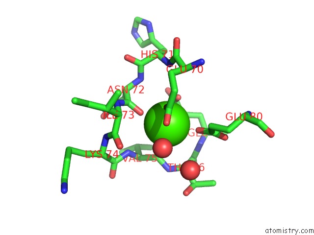
Mono view
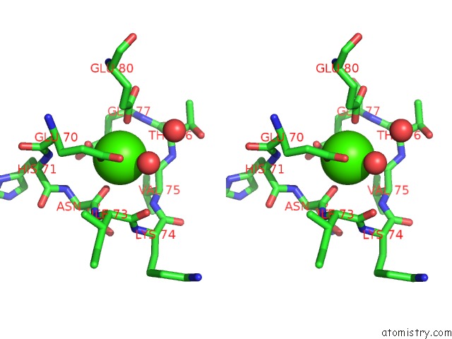
Stereo pair view
Mono view
Stereo pair view
A full contact list of Calcium with other atoms in the Ca binding
site number 1 of Anionic Salmon Trypsin in Complex with Squash Seed Inhibitor (Cucurbita Pepo Trypsin Inhibitor II) within 5.0Å range:
|
Reference:
R.Helland,
G.I.Berglund,
J.Otlewski,
W.Apostoluk,
O.A.Andersen,
N.P.Willassen,
A.O.Smalas.
High-Resolution Structures of Three New Trypsin-Squash-Inhibitor Complexes: A Detailed Comparison with Other Trypsins and Their Complexes. Acta Crystallogr.,Sect.D V. 55 139 1999.
ISSN: ISSN 0907-4449
PubMed: 10089404
DOI: 10.1107/S090744499801052X
Page generated: Tue Jul 8 08:22:49 2025
ISSN: ISSN 0907-4449
PubMed: 10089404
DOI: 10.1107/S090744499801052X
Last articles
Mg in 7JGRMg in 7JGA
Mg in 7JG5
Mg in 7JFP
Mg in 7GYI
Mg in 7GYH
Mg in 7GYG
Mg in 7GYF
Mg in 7GYE
Mg in 7GYD