Calcium »
PDB 4a42-4adj »
4a4b »
Calcium in PDB 4a4b: Structure of Modified PHOSPHOTYR371-C-Cbl-UBCH5B-Zap-70 Complex
Enzymatic activity of Structure of Modified PHOSPHOTYR371-C-Cbl-UBCH5B-Zap-70 Complex
All present enzymatic activity of Structure of Modified PHOSPHOTYR371-C-Cbl-UBCH5B-Zap-70 Complex:
2.7.10.2; 6.3.2.19;
2.7.10.2; 6.3.2.19;
Protein crystallography data
The structure of Structure of Modified PHOSPHOTYR371-C-Cbl-UBCH5B-Zap-70 Complex, PDB code: 4a4b
was solved by
H.Dou,
L.Buetow,
A.Hock,
G.J.Sibbet,
K.H.Vousden,
D.T.Huang,
with X-Ray Crystallography technique. A brief refinement statistics is given in the table below:
| Resolution Low / High (Å) | 28.725 / 2.79 |
| Space group | P 65 2 2 |
| Cell size a, b, c (Å), α, β, γ (°) | 74.112, 74.112, 449.706, 90.00, 90.00, 120.00 |
| R / Rfree (%) | 18.74 / 26.68 |
Other elements in 4a4b:
The structure of Structure of Modified PHOSPHOTYR371-C-Cbl-UBCH5B-Zap-70 Complex also contains other interesting chemical elements:
| Zinc | (Zn) | 2 atoms |
Calcium Binding Sites:
The binding sites of Calcium atom in the Structure of Modified PHOSPHOTYR371-C-Cbl-UBCH5B-Zap-70 Complex
(pdb code 4a4b). This binding sites where shown within
5.0 Angstroms radius around Calcium atom.
In total only one binding site of Calcium was determined in the Structure of Modified PHOSPHOTYR371-C-Cbl-UBCH5B-Zap-70 Complex, PDB code: 4a4b:
In total only one binding site of Calcium was determined in the Structure of Modified PHOSPHOTYR371-C-Cbl-UBCH5B-Zap-70 Complex, PDB code: 4a4b:
Calcium binding site 1 out of 1 in 4a4b
Go back to
Calcium binding site 1 out
of 1 in the Structure of Modified PHOSPHOTYR371-C-Cbl-UBCH5B-Zap-70 Complex
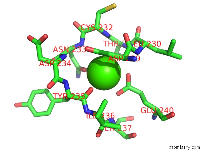
Mono view
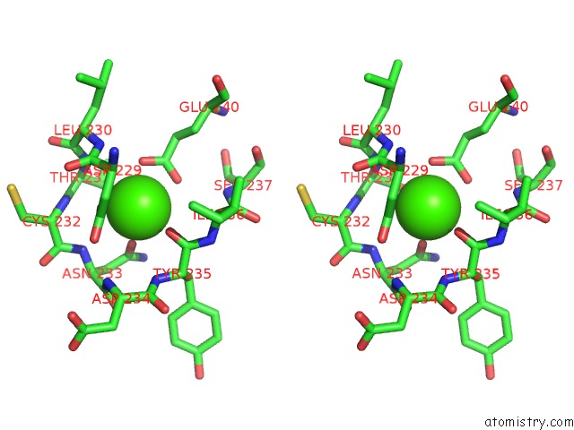
Stereo pair view
Mono view
Stereo pair view
A full contact list of Calcium with other atoms in the Ca binding
site number 1 of Structure of Modified PHOSPHOTYR371-C-Cbl-UBCH5B-Zap-70 Complex within 5.0Å range:
|
Reference:
H.Dou,
L.Buetow,
A.Hock,
G.J.Sibbet,
K.H.Vousden,
D.T.Huang.
Structural Basis For Autoinhibition and Phosphorylation-Dependent Activation of C-Cbl Nat.Struct.Mol.Biol. V. 19 184 2012.
ISSN: ISSN 1545-9993
PubMed: 22266821
DOI: 10.1038/NSMB.2231
Page generated: Tue Jul 8 18:24:29 2025
ISSN: ISSN 1545-9993
PubMed: 22266821
DOI: 10.1038/NSMB.2231
Last articles
Mg in 6IGJMg in 6IG0
Mg in 6IFR
Mg in 6IFK
Mg in 6ICZ
Mg in 6IFH
Mg in 6ID1
Mg in 6IFD
Mg in 6IF7
Mg in 6ID0