Calcium »
PDB 4ry0-4twe »
4sgb »
Calcium in PDB 4sgb: Structure of the Complex of Streptomyces Griseus Proteinase B and Polypeptide Chymotrypsin Inhibitor-1 From Russet Burbank Potato Tubers at 2.1 Angstroms Resolution
Protein crystallography data
The structure of Structure of the Complex of Streptomyces Griseus Proteinase B and Polypeptide Chymotrypsin Inhibitor-1 From Russet Burbank Potato Tubers at 2.1 Angstroms Resolution, PDB code: 4sgb
was solved by
M.James,
H.Greenblatt,
with X-Ray Crystallography technique. A brief refinement statistics is given in the table below:
| Resolution Low / High (Å) | 8.00 / 2.10 |
| Space group | P 1 21 1 |
| Cell size a, b, c (Å), α, β, γ (°) | 50.920, 46.200, 52.530, 90.00, 117.08, 90.00 |
| R / Rfree (%) | n/a / n/a |
Calcium Binding Sites:
The binding sites of Calcium atom in the Structure of the Complex of Streptomyces Griseus Proteinase B and Polypeptide Chymotrypsin Inhibitor-1 From Russet Burbank Potato Tubers at 2.1 Angstroms Resolution
(pdb code 4sgb). This binding sites where shown within
5.0 Angstroms radius around Calcium atom.
In total only one binding site of Calcium was determined in the Structure of the Complex of Streptomyces Griseus Proteinase B and Polypeptide Chymotrypsin Inhibitor-1 From Russet Burbank Potato Tubers at 2.1 Angstroms Resolution, PDB code: 4sgb:
In total only one binding site of Calcium was determined in the Structure of the Complex of Streptomyces Griseus Proteinase B and Polypeptide Chymotrypsin Inhibitor-1 From Russet Burbank Potato Tubers at 2.1 Angstroms Resolution, PDB code: 4sgb:
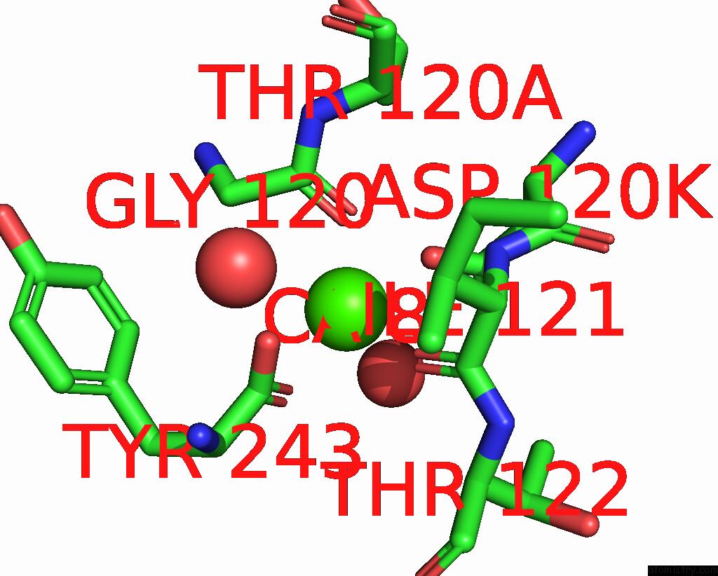
Calcium binding site 1 out of 1 in 4sgb
Go back to
Calcium binding site 1 out
of 1 in the Structure of the Complex of Streptomyces Griseus Proteinase B and Polypeptide Chymotrypsin Inhibitor-1 From Russet Burbank Potato Tubers at 2.1 Angstroms Resolution
Mono view

Stereo pair view
Mono view
Stereo pair view
A full contact list of Calcium with other atoms in the Ca binding
site number 1 of Structure of the Complex of Streptomyces Griseus Proteinase B and Polypeptide Chymotrypsin Inhibitor-1 From Russet Burbank Potato Tubers at 2.1 Angstroms Resolution within 5.0Å range:
|
Reference:
H.M.Greenblatt,
C.A.Ryan,
M.N.James.
Structure of the Complex of Streptomyces Griseus Proteinase B and Polypeptide Chymotrypsin Inhibitor-1 From Russet Burbank Potato Tubers at 2.1 A Resolution. J.Mol.Biol. V. 205 201 1989.
ISSN: ISSN 0022-2836
PubMed: 2494344
DOI: 10.1016/0022-2836(89)90376-8
Page generated: Wed Jul 9 02:03:51 2025
ISSN: ISSN 0022-2836
PubMed: 2494344
DOI: 10.1016/0022-2836(89)90376-8
Last articles
Mg in 6P1ZMg in 6P22
Mg in 6P1W
Mg in 6P1T
Mg in 6P1V
Mg in 6P1X
Mg in 6P1R
Mg in 6P1Q
Mg in 6P1P
Mg in 6P1L