Calcium »
PDB 8deo-8e4m »
8dvy »
Calcium in PDB 8dvy: Dna Glycosylase Muty Variant N146S in Complex with Dna Containing D(8- Oxo-G) Paired with An Enzyme-Generated Abasic Site Product (Ap) and Crystalized with Calcium Acetate
Enzymatic activity of Dna Glycosylase Muty Variant N146S in Complex with Dna Containing D(8- Oxo-G) Paired with An Enzyme-Generated Abasic Site Product (Ap) and Crystalized with Calcium Acetate
All present enzymatic activity of Dna Glycosylase Muty Variant N146S in Complex with Dna Containing D(8- Oxo-G) Paired with An Enzyme-Generated Abasic Site Product (Ap) and Crystalized with Calcium Acetate:
3.2.2.31;
3.2.2.31;
Protein crystallography data
The structure of Dna Glycosylase Muty Variant N146S in Complex with Dna Containing D(8- Oxo-G) Paired with An Enzyme-Generated Abasic Site Product (Ap) and Crystalized with Calcium Acetate, PDB code: 8dvy
was solved by
M.Demir,
L.P.Russelburg,
M.P.Horvath,
S.S.David,
with X-Ray Crystallography technique. A brief refinement statistics is given in the table below:
| Resolution Low / High (Å) | 70.35 / 2.36 |
| Space group | P 21 21 21 |
| Cell size a, b, c (Å), α, β, γ (°) | 37.86, 85.48, 140.71, 90, 90, 90 |
| R / Rfree (%) | 24.7 / 27.4 |
Other elements in 8dvy:
The structure of Dna Glycosylase Muty Variant N146S in Complex with Dna Containing D(8- Oxo-G) Paired with An Enzyme-Generated Abasic Site Product (Ap) and Crystalized with Calcium Acetate also contains other interesting chemical elements:
| Iron | (Fe) | 4 atoms |
Calcium Binding Sites:
The binding sites of Calcium atom in the Dna Glycosylase Muty Variant N146S in Complex with Dna Containing D(8- Oxo-G) Paired with An Enzyme-Generated Abasic Site Product (Ap) and Crystalized with Calcium Acetate
(pdb code 8dvy). This binding sites where shown within
5.0 Angstroms radius around Calcium atom.
In total 3 binding sites of Calcium where determined in the Dna Glycosylase Muty Variant N146S in Complex with Dna Containing D(8- Oxo-G) Paired with An Enzyme-Generated Abasic Site Product (Ap) and Crystalized with Calcium Acetate, PDB code: 8dvy:
Jump to Calcium binding site number: 1; 2; 3;
In total 3 binding sites of Calcium where determined in the Dna Glycosylase Muty Variant N146S in Complex with Dna Containing D(8- Oxo-G) Paired with An Enzyme-Generated Abasic Site Product (Ap) and Crystalized with Calcium Acetate, PDB code: 8dvy:
Jump to Calcium binding site number: 1; 2; 3;
Calcium binding site 1 out of 3 in 8dvy
Go back to
Calcium binding site 1 out
of 3 in the Dna Glycosylase Muty Variant N146S in Complex with Dna Containing D(8- Oxo-G) Paired with An Enzyme-Generated Abasic Site Product (Ap) and Crystalized with Calcium Acetate
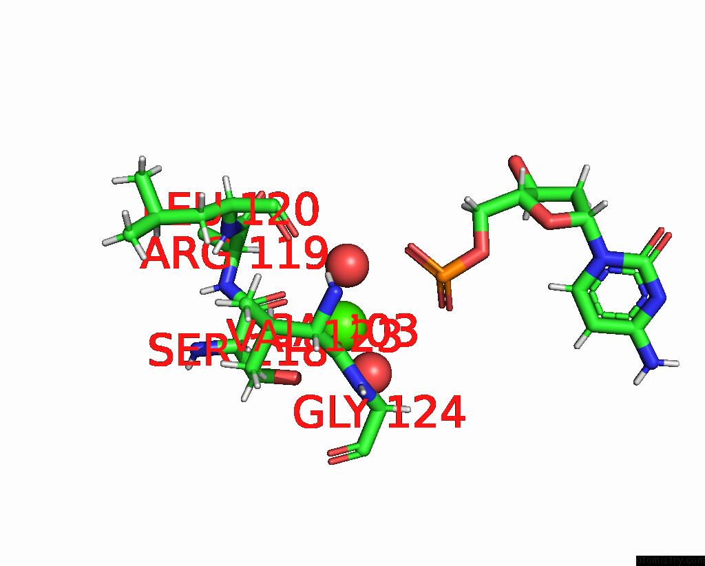
Mono view

Stereo pair view
Mono view
Stereo pair view
A full contact list of Calcium with other atoms in the Ca binding
site number 1 of Dna Glycosylase Muty Variant N146S in Complex with Dna Containing D(8- Oxo-G) Paired with An Enzyme-Generated Abasic Site Product (Ap) and Crystalized with Calcium Acetate within 5.0Å range:
|
Calcium binding site 2 out of 3 in 8dvy
Go back to
Calcium binding site 2 out
of 3 in the Dna Glycosylase Muty Variant N146S in Complex with Dna Containing D(8- Oxo-G) Paired with An Enzyme-Generated Abasic Site Product (Ap) and Crystalized with Calcium Acetate
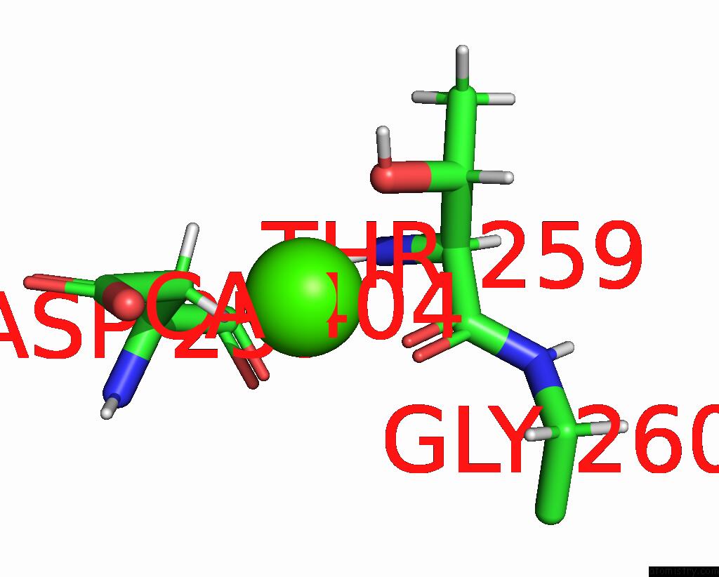
Mono view
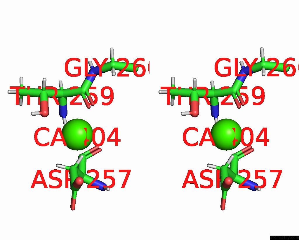
Stereo pair view
Mono view
Stereo pair view
A full contact list of Calcium with other atoms in the Ca binding
site number 2 of Dna Glycosylase Muty Variant N146S in Complex with Dna Containing D(8- Oxo-G) Paired with An Enzyme-Generated Abasic Site Product (Ap) and Crystalized with Calcium Acetate within 5.0Å range:
|
Calcium binding site 3 out of 3 in 8dvy
Go back to
Calcium binding site 3 out
of 3 in the Dna Glycosylase Muty Variant N146S in Complex with Dna Containing D(8- Oxo-G) Paired with An Enzyme-Generated Abasic Site Product (Ap) and Crystalized with Calcium Acetate
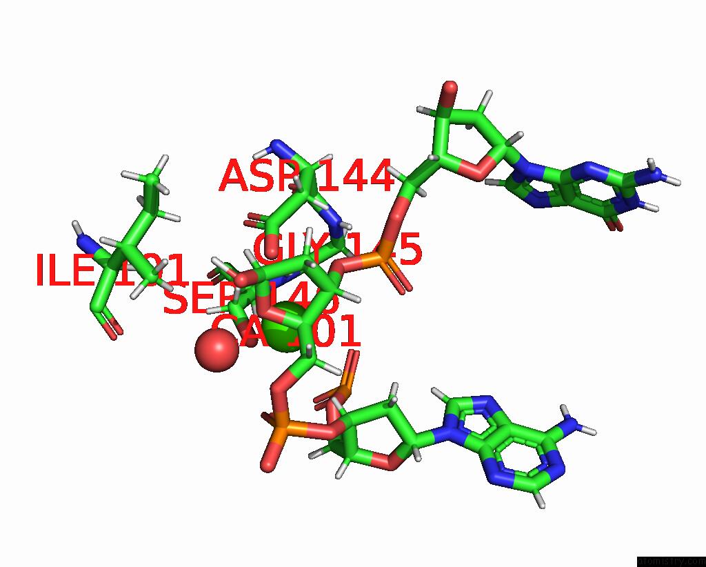
Mono view

Stereo pair view
Mono view
Stereo pair view
A full contact list of Calcium with other atoms in the Ca binding
site number 3 of Dna Glycosylase Muty Variant N146S in Complex with Dna Containing D(8- Oxo-G) Paired with An Enzyme-Generated Abasic Site Product (Ap) and Crystalized with Calcium Acetate within 5.0Å range:
|
Reference:
M.Demir,
L.P.Russelburg,
J.Lin,
C.H.Trasvina-Arenas,
B.Huang,
P.K.Yuen,
M.P.Horvath,
S.S.David.
Structural Snapshots of Base Excision Mechanism By Dna Glycosylase Muty Reveal Retention of Stereochemistry For Abasic Site Product Formation To Be Published.
Page generated: Thu Jul 10 04:00:29 2025
Last articles
Na in 2QB4Na in 2QD6
Na in 2QCI
Na in 2QA0
Na in 2Q95
Na in 2Q96
Na in 2Q94
Na in 2Q93
Na in 2Q8X
Na in 2Q72