Calcium »
PDB 9ezl-9fzc »
9f1z »
Calcium in PDB 9f1z: Crystal Structure of Adenylyate Cyclase Variant Y6W
Protein crystallography data
The structure of Crystal Structure of Adenylyate Cyclase Variant Y6W, PDB code: 9f1z
was solved by
S.Kapetanaki,
N.Coquelle,
M.Weik,
with X-Ray Crystallography technique. A brief refinement statistics is given in the table below:
| Resolution Low / High (Å) | 46.66 / 1.95 |
| Space group | P 21 21 2 |
| Cell size a, b, c (Å), α, β, γ (°) | 100.455, 52.684, 71.525, 90, 90, 90 |
| R / Rfree (%) | 19.6 / 23.4 |
Calcium Binding Sites:
The binding sites of Calcium atom in the Crystal Structure of Adenylyate Cyclase Variant Y6W
(pdb code 9f1z). This binding sites where shown within
5.0 Angstroms radius around Calcium atom.
In total 2 binding sites of Calcium where determined in the Crystal Structure of Adenylyate Cyclase Variant Y6W, PDB code: 9f1z:
Jump to Calcium binding site number: 1; 2;
In total 2 binding sites of Calcium where determined in the Crystal Structure of Adenylyate Cyclase Variant Y6W, PDB code: 9f1z:
Jump to Calcium binding site number: 1; 2;
Calcium binding site 1 out of 2 in 9f1z
Go back to
Calcium binding site 1 out
of 2 in the Crystal Structure of Adenylyate Cyclase Variant Y6W
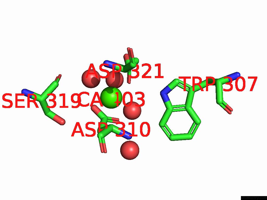
Mono view
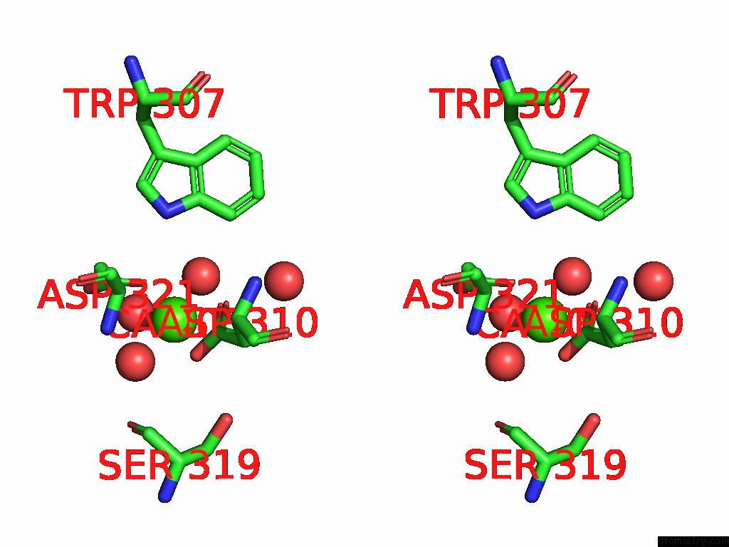
Stereo pair view
Mono view
Stereo pair view
A full contact list of Calcium with other atoms in the Ca binding
site number 1 of Crystal Structure of Adenylyate Cyclase Variant Y6W within 5.0Å range:
|
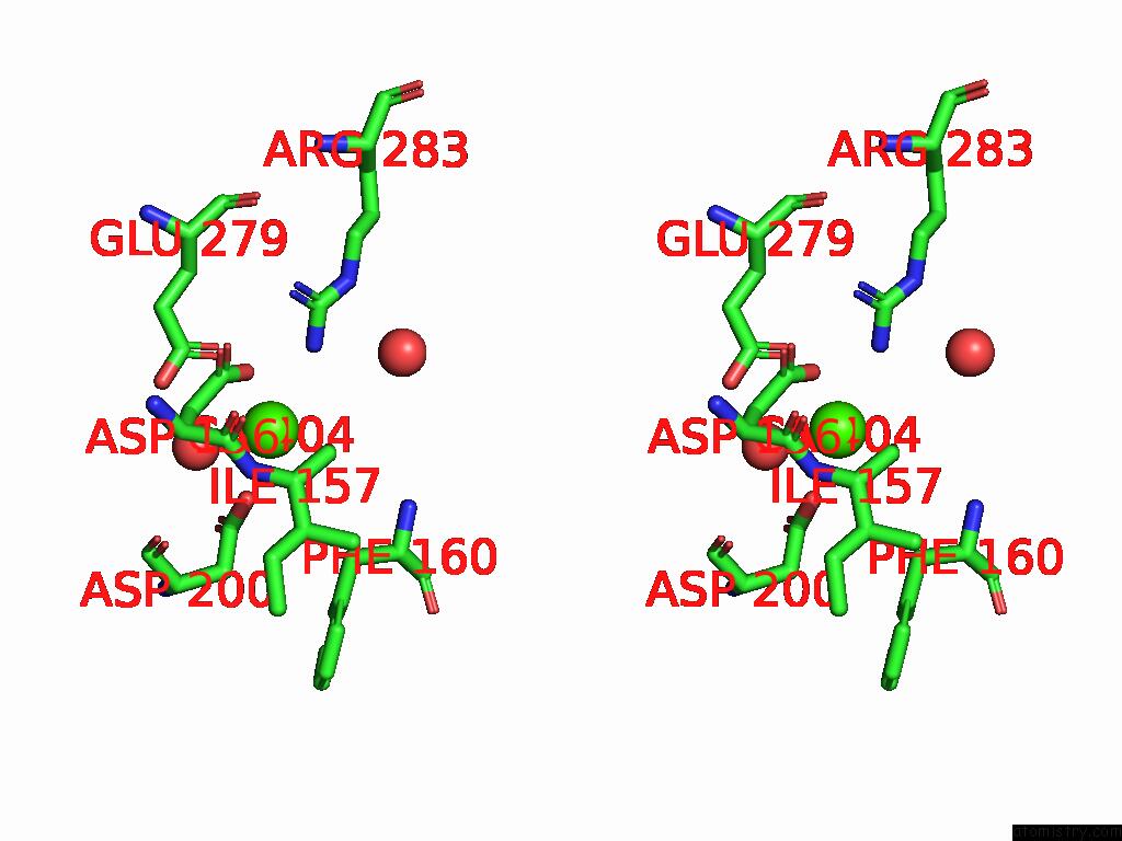
Calcium binding site 2 out of 2 in 9f1z
Go back to
Calcium binding site 2 out
of 2 in the Crystal Structure of Adenylyate Cyclase Variant Y6W
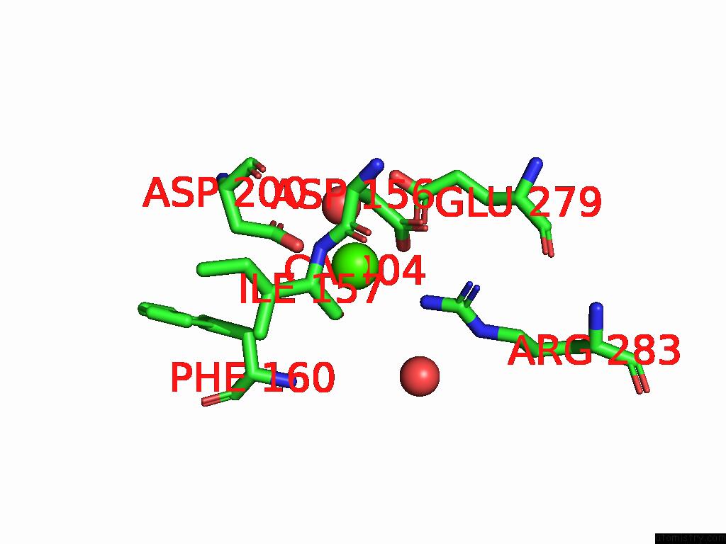
Mono view
Stereo pair view
Mono view
Stereo pair view
A full contact list of Calcium with other atoms in the Ca binding
site number 2 of Crystal Structure of Adenylyate Cyclase Variant Y6W within 5.0Å range:
|
Reference:
S.Kapetanaki,
N.Coquelle,
M.Weik.
Sfx Room Temperature Structure of Oscillatoria Acuminata Adenylyate Cyclase. To Be Published.
Page generated: Thu Jul 10 09:37:47 2025
Last articles
Fe in 2YXOFe in 2YRS
Fe in 2YXC
Fe in 2YNM
Fe in 2YVJ
Fe in 2YP1
Fe in 2YU2
Fe in 2YU1
Fe in 2YQB
Fe in 2YOO