Calcium »
PDB 1scr-1spj »
1sgt »
Calcium in PDB 1sgt: Refined Crystal Structure of Streptomyces Griseus Trypsin at 1.7 Angstroms Resolution
Enzymatic activity of Refined Crystal Structure of Streptomyces Griseus Trypsin at 1.7 Angstroms Resolution
All present enzymatic activity of Refined Crystal Structure of Streptomyces Griseus Trypsin at 1.7 Angstroms Resolution:
3.4.21.4;
3.4.21.4;
Protein crystallography data
The structure of Refined Crystal Structure of Streptomyces Griseus Trypsin at 1.7 Angstroms Resolution, PDB code: 1sgt
was solved by
R.J.Read,
M.N.G.James,
with X-Ray Crystallography technique. A brief refinement statistics is given in the table below:
| Resolution Low / High (Å) | 8.00 / 1.70 |
| Space group | C 2 2 21 |
| Cell size a, b, c (Å), α, β, γ (°) | 72.290, 50.980, 120.090, 90.00, 90.00, 90.00 |
| R / Rfree (%) | n/a / n/a |
Calcium Binding Sites:
The binding sites of Calcium atom in the Refined Crystal Structure of Streptomyces Griseus Trypsin at 1.7 Angstroms Resolution
(pdb code 1sgt). This binding sites where shown within
5.0 Angstroms radius around Calcium atom.
In total only one binding site of Calcium was determined in the Refined Crystal Structure of Streptomyces Griseus Trypsin at 1.7 Angstroms Resolution, PDB code: 1sgt:
In total only one binding site of Calcium was determined in the Refined Crystal Structure of Streptomyces Griseus Trypsin at 1.7 Angstroms Resolution, PDB code: 1sgt:
Calcium binding site 1 out of 1 in 1sgt
Go back to
Calcium binding site 1 out
of 1 in the Refined Crystal Structure of Streptomyces Griseus Trypsin at 1.7 Angstroms Resolution
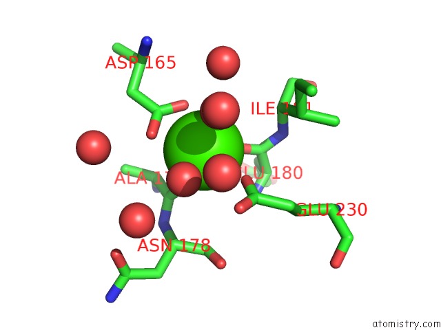
Mono view
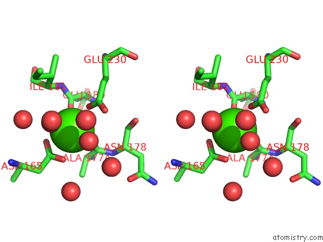
Stereo pair view
Mono view
Stereo pair view
A full contact list of Calcium with other atoms in the Ca binding
site number 1 of Refined Crystal Structure of Streptomyces Griseus Trypsin at 1.7 Angstroms Resolution within 5.0Å range:
|
Reference:
R.J.Read,
M.N.James.
Refined Crystal Structure of Streptomyces Griseus Trypsin at 1.7 A Resolution. J.Mol.Biol. V. 200 523 1988.
ISSN: ISSN 0022-2836
PubMed: 3135412
DOI: 10.1016/0022-2836(88)90541-4
Page generated: Tue Jul 8 01:56:32 2025
ISSN: ISSN 0022-2836
PubMed: 3135412
DOI: 10.1016/0022-2836(88)90541-4
Last articles
Ca in 2I44Ca in 2I4C
Ca in 2HYW
Ca in 2I4B
Ca in 2I3Q
Ca in 2I3P
Ca in 2I1Q
Ca in 2I1P
Ca in 2I0U
Ca in 2I0F